

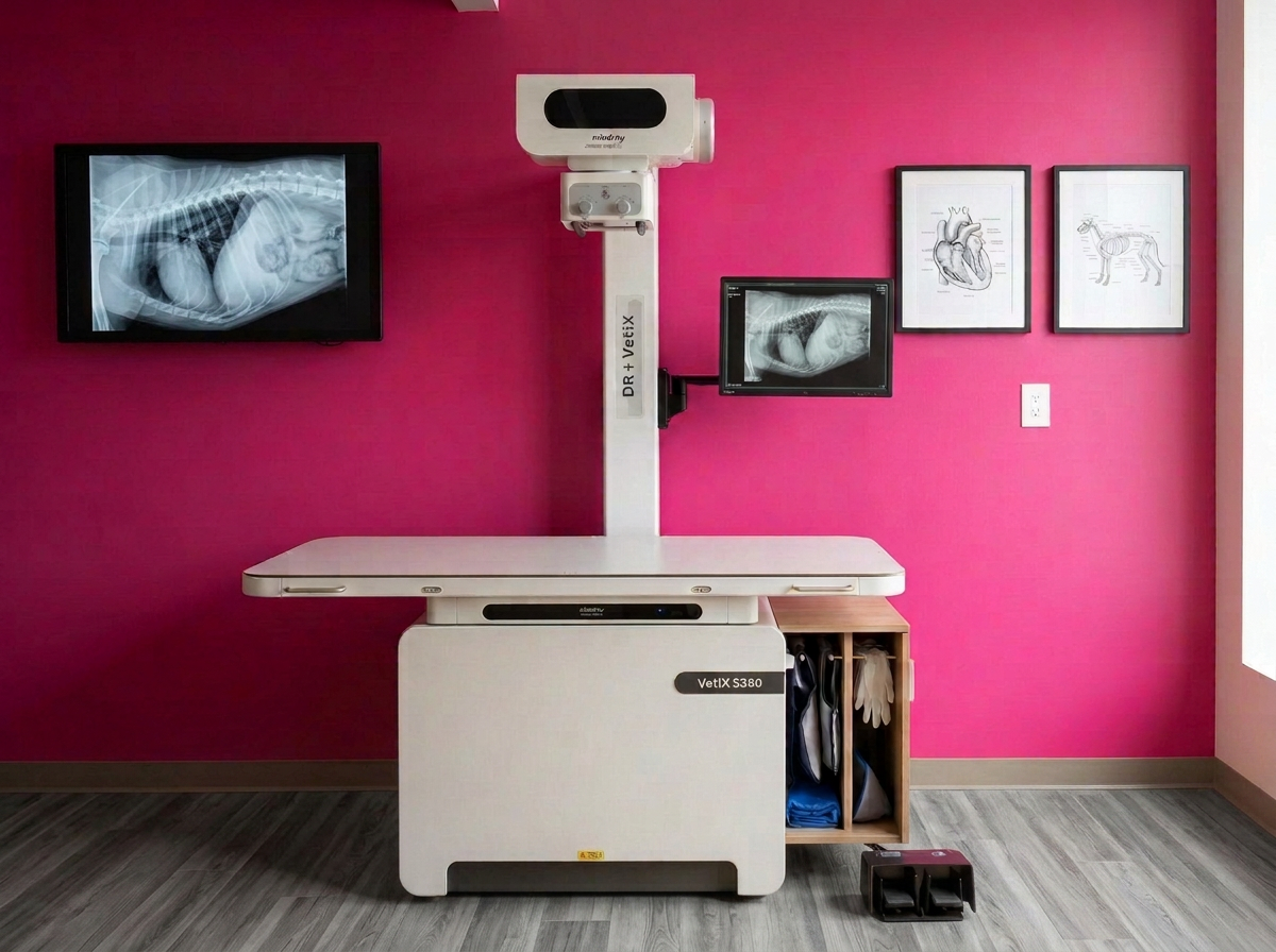
Advanced diagnostic imaging to evaluate your pet’s heart, lungs, and thoracic health.
Radiographs, commonly known as X-rays, are one of the most vital, non-invasive diagnostic tools used in veterinary cardiology. Digital X-rays allow our veterinary cardiologist to immediately capture detailed, high-resolution images of your dog or cat’s chest, providing critical details about heart size, shape, and surrounding lung tissue.
At TriState Cardiology for Pets, digital radiography provides near-instant results so we can quickly identify underlying cardiac conditions, evaluate respiratory symptoms, and create a tailored treatment plan for your pet.
Digital Radiography (X-Ray) Services
A digital X-ray (radiograph) is a quick, non-invasive imaging technique that uses low-dose radiation to produce clear, instantaneous pictures of your pet’s internal structures. Unlike traditional film X-rays, digital radiography yields high-definition images in a matter of seconds, allowing for immediate analysis during your pet’s appointment.
In veterinary cardiology, chest radiographs (thoracic X-rays) are particularly essential. They allow our specialty team to examine:
Heart Size & Silhouette: Assessing overall cardiac enlargement or abnormalities in heart shape.
Lung Fields & Airways: Checking for fluid accumulation, inflammation, or air space disease in the lungs.
Pulmonary Vessels: Evaluating blood vessels leading to and from the heart for signs of hypertension or blockage.
Pleural Space & Structures: Identifying fluid or air around the lungs and examining surrounding bony structures.
Why Would My Dog or Cat Need a Digital X-Ray?
Your primary veterinarian or veterinary cardiologist may recommend chest digital X-rays if your pet displays signs of heart or respiratory distress. X-rays are a foundational diagnostic step for pets exhibiting:
Persistent coughing or gagging
Rapid, labored, or difficult breathing
Unexplained fatigue or exercise intolerance
Heart murmurs or abnormal heart rhythms detected during a physical exam
Fainting spells or sudden collapse episodes
How Digital X-Rays Help Diagnose Congestive Heart Failure
Chest X-rays are essential for diagnosing and managing Congestive Heart Failure (CHF) in dogs and cats. While an echocardiogram assesses heart function and valve movement, X-rays are the primary tool used to detect fluid buildup in the lungs (pulmonary edema).
A diagnosis of CHF is confirmed when digital radiographs show evidence of pulmonary fluid accumulation alongside cardiac enlarged structure changes. Because digital results are ready within seconds, our team can begin life-saving stabilization and medical therapy immediately.
What Happens During a Digital Radiography Exam?
Digital X-rays are non-invasive and painless. Your pet is gently positioned on a padded X-ray table while our team takes two or three view angles of the chest.
Fast & Gentle: The imaging process takes only a few minutes, minimizing stress for pets with breathing difficulties.
Minimal Restraint: Most pets tolerate the procedure easily without the need for sedation or anesthesia.
Instant Digital Quality: Images appear on our diagnostic monitors within seconds, allowing for instant review and consultation.
What Is a Digital Veterinary X-Ray?
Digital X-Ray FAQs
-
A digital X-ray provides a static image of the overall size and shape of the heart and evaluates whether fluid is building up in the lungs. An echocardiogram uses ultrasound to look inside the beating heart to assess valve movement, chamber wall thickness, and blood flow velocity. Both tests complement each other to give a complete cardiac picture.
-
Yes. Digital radiography uses extremely low levels of radiation, making it very safe for pets. Because digital sensors are far more sensitive than traditional film, exposures are shorter and fewer retakes are needed.
-
In most cases, sedation is not necessary. The digital imaging process takes only seconds, so gentle physical handling is usually all that is required. If a pet is extremely anxious or in severe pain from an unrelated condition, light sedation may be discussed to ensure comfort.
-
Results are available within seconds. Digital technology allows our cardiologist to analyze the images immediately during your appointment and discuss the diagnosis and care plan with you right away.
-
Yes. Chest radiographs are the gold standard for identifying fluid in the lung tissue (pulmonary edema) or around the lungs (pleural effusion), which are key markers for congestive heart failure and respiratory conditions.