

Echocardiogram & Diagnostics
Advanced ultrasound imaging to evaluate your pet’s heart
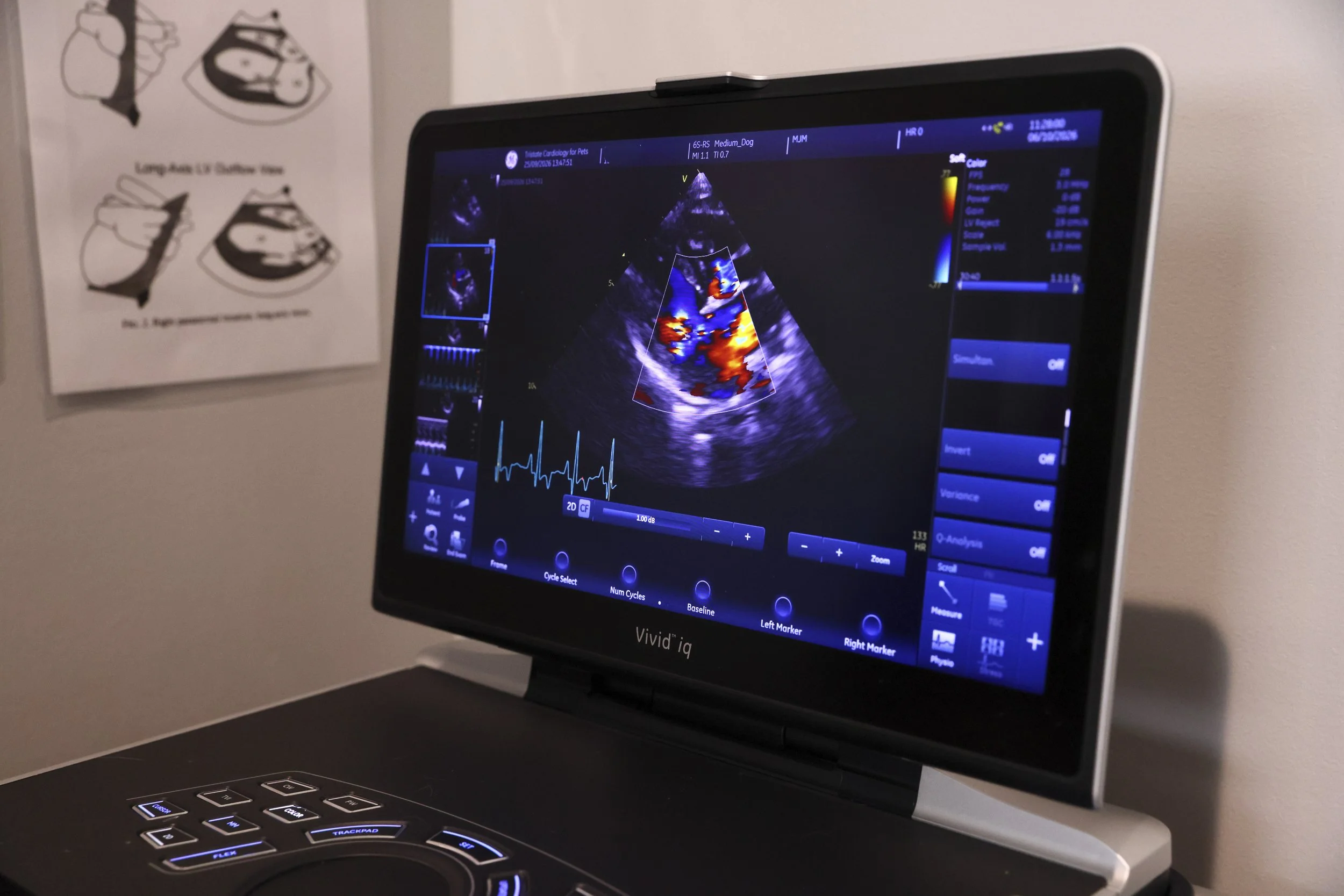
An echocardiogram is a non-invasive ultrasound examination of the heart that allows a veterinary cardiologist to see how your dog or cat’s heart is working in real time. Unlike standard X-rays, which primarily show the size of the heart and the surrounding lungs, an echocardiogram provides detailed information about the heart muscle, chambers, valves, and blood flow.
At TriState Cardiology for Pets, echocardiography helps our doctor identify and evaluate heart disease so she can develop an appropriate, individualized treatment plan for your pet.
An echocardiogram, sometimes called a cardiac ultrasound, uses sound waves to create moving images of the heart. It allows the veterinary cardiologist to evaluate both the structure and function of your pet’s heart.
During the examination, the cardiologist can assess:
How well the heart muscle contracts and relaxes
The size and thickness of the heart chambers and walls
Blood flow through the heart and major blood vessels
The function of the heart valves
Valve leakage and abnormal blood flow
Fluid surrounding the heart
Abnormal masses or other changes
Other structural or functional abnormalities associated with heart disease
Because the heart can be observed while it is beating, an echocardiogram provides information that cannot be obtained from an X-ray alone.
Why Would My Dog or Cat Need an Echocardiogram?
Your veterinarian may recommend an echocardiogram if your pet has a heart murmur, abnormal heart rhythm, breathing concerns, exercise intolerance, fainting episodes, or other signs that could indicate heart disease.
An echocardiogram can help determine whether heart disease is present, what type of heart disease your pet has, and how severe it is.
The results can also help the veterinary cardiologist determine whether treatment is needed and which treatment approach is most appropriate.
What Happens During an Echocardiogram?
An echocardiogram is non-invasive and generally not painful.
Your pet will lie on a padded examination table while the cardiologist performs the ultrasound examination. A small area of hair may need to be clipped so the ultrasound probe can make good contact with the skin.
The probe is placed against the chest over the heart, allowing the cardiologist to obtain detailed images of the heart as it beats.
Most echocardiogram examinations take approximately 15–25 minutes, although the exact time can vary depending on your pet and the complexity of the examination.
What Can an Echocardiogram Tell Us About My Pet's Heart?
An echocardiogram provides detailed information about both the structure and function of the heart.
It can help the cardiologist determine the location and type of cardiac abnormality and assess its severity. This information is important when deciding whether your pet needs medication, monitoring, additional testing, or another form of treatment.
Most importantly, the information obtained from an echocardiogram helps your veterinary cardiologist create a treatment plan tailored to your pet while keeping quality of life at the center of care.
What Is an Echocardiogram?
Echocardiogram FAQs
-
No. An echocardiogram is a non-invasive ultrasound examination and is generally not painful. Your pet simply needs to remain in position while the ultrasound probe is placed against the chest.
-
Most echocardiograms take approximately 15–25 minutes. More complex cases may require additional time for a thorough cardiac evaluation.
-
Many dogs and cats can have an echocardiogram without sedation. Whether sedation is appropriate depends on your pet’s temperament, medical condition, and ability to remain comfortably positioned during the examination.
-
An X-ray primarily provides an image of the heart's overall size and the surrounding structures, including the lungs. An echocardiogram uses ultrasound to produce moving images of the heart, allowing the cardiologist to evaluate heart muscle movement, chamber dimensions, valves, and blood flow.
-
X-rays and echocardiograms provide different types of information. X-rays can help evaluate heart size and the lungs, while an echocardiogram provides a much more detailed assessment of the heart's structure and function. Your veterinarian or cardiologist may use both tests because they complement one another.scription
-
Yes. Echocardiography is an important diagnostic tool for identifying and evaluating many types of heart disease in dogs and cats. It can show abnormalities involving the heart muscle, chambers, valves, blood flow, and other cardiac structures.
-
An echocardiogram can help identify abnormalities such as changes in heart chamber size, thickened heart walls, abnormal valve function or leakage, changes in heart muscle contraction, abnormal blood flow, fluid around the heart, and certain masses or clots.
-
Not necessarily. The echocardiogram is a diagnostic test that helps determine whether heart disease is present and how significant it is. Depending on the findings, your cardiologist may recommend treatment, monitoring, additional testing, or no immediate treatment.
-
Not necessarily. An echocardiogram is commonly recommended when a veterinarian wants more information about a pet's heart. It provides detailed information that can help determine whether a heart problem is significant and what, if anything, should be done about it.